#apaperaday: Structural and Ultrastructural Changes in the Tongue of mdx Mice
In today’s #apaperaday, Prof. Aartsma-Rus reads and comments on the paper titled: Structural and Ultrastructural Changes in the Tongue of mdx Mice.
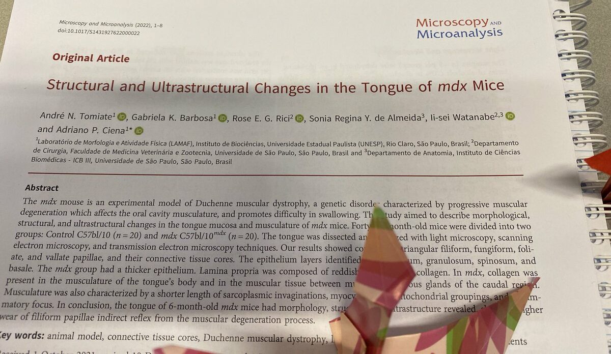
Today’s pick is from Tomiate et al from the journal microscopy and microanalysis on the changes in the tongue of 6 months old mdx mice. Doi 10.1017/s1431927622000022.
Due to lack of dystrophin, skeletal muscles of Duchenne patients deteriorate. This also affects the masseter and tongue muscles (required for chewing and swallowing). In older Duchenne patients this results in problems with food intake and swallowing difficulties.
Authors here do a detailed analysis of the tongue of mdx mice – a mouse model for Duchenne. They used 20 mdx and 20 wild type mice for detailed microscopy. It is not clear why they needed this many animals as for most analyses work is descriptive and images from N=1 are shown.
Images are beautiful though. Authors find that the epithelium of mdx mouse tongue is thicker, that there is inflammation and extra collagen deposition. Papillae look normal. Disorganization of mitochondria was observed and mdx mice have less serous glands and more mucous glands.

One more picture from a scanning electron microscope because it is beautiful. Authors discuss that there are signs of muscular dystrophy and fibrosis present at 6 months. Earlier work showed no difference in 100 day old mdx mice, while 26 months old mice are severely fibrotic

Authors conclude atrophy and fibrosis deposition occurs mostly between 6 and 26 months in mice. I like the beautiful pictures and I understand the focus was on microscopy. However, a form vs function combination would have been better – how was function impacted in these mice?
Knowing only histology/form tells us just one part of the story. Also would like to see different timepoints to see how the pathology develops. So despite the beautiful images, I am a bit disappointed.
Pictures by Annemieke, used with permission.